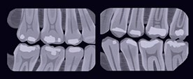


Checkup dental radiographs, or X-rays, are a commonality at your dental visit. Bite-wings radiographs are an essential screening assessment needed to diagnose dental cavities that form between the teeth and are undetectable to the human eye. A full set of radiographs is taken once every 3-5 years to evaluate the roots of all the teeth and rule out unwanted pathology/infection. These conventional radiographs are necessary to complete a comprehensive exam on a patient, but these radiographs cannot tell us everything that is going on with a patient’s health.
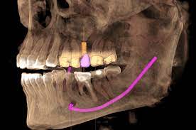
Cone beam, or CBCT scans have been introduced to the dental world and have been around for years now. What makes this scan different from conventional radiographs mentioned above is that it provides a reproduced 3-demensional image of the head and neck, whereas standard radiographs produce a 2-demensiaonal image of the teeth. A cone beam scan produces a high-definition radiograph that picks up infection/cavitations in its early stages, when the standard radiograph could miss one. Cone beam scans are becoming a standard assessment and should be especially utilized when evaluating a patient with an extensive history of root canals, implants, and crowns. Faulty dental work that has been in the mouth for many years tends to allow infection to start in the surrounding bone that would otherwise go undetected without a cone beam scan.
Not only can a cone beam scan detect pathology, but it can also help us to evaluate the jaw joints (TMJ), sinus, and airway health. Patients undergoing a sleep study can benefit largely from a cone beam scan evaluation as it can allow us to see if the airway is abnormally small. Breathing issues may not be related to the airway alone as a deviated nasal septum or other sinus issues can be the main culprit. If there is anything unfavorable going on in the nose, the cone beam scan will be able identify the problem. Patients considered for tongue tie release should have a cone beam scan done as it shows exact positioning of the tongue in relationship to its surrounding anatomy. Some patients may not undergo togue tie release based on the findings of their scan. However, A cone beam scan’s diagnostic ability does not stop here. There are various uses that come from this assessment, and it will soon become common practice at the dental office.